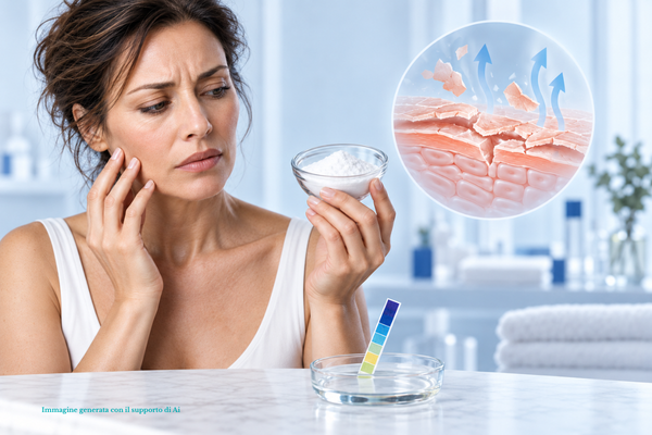



Hyperpigmentation appears as areas of the face with an uneven complexion, with darker patches than the rest of the skin tone. The common term is "spot", but in dermatology, hyperpigmentation refers to a broader and more complex process.
The distinction is not a theoretical exercise: it is the key to identifying the exact cause and adopting the right strategy. The same blemish can in fact conceal different mechanisms — hormonal, post-inflammatory or sun-induced.
Article written by Elisa Avalle, founder of LeLang Skin Care and dermocosmetics specialist. Cover image generated with the support of artificial intelligence.
[INDICE:h2]
Hyperpigmentation: not just "spots"
It is a condition characterized by excessive synthesis and localized accumulation of melanin, the pigment produced by melanocytes.
When the skin is subjected to a stimulus — UV rays, inflammation or hormonal changes — melanocytes produce an excess of pigment that deposits in the epidermis or dermis.
The difference between the two lies here: hyperpigmentation is the underlying biological mechanism, while the spot is the visible result.
Treating hyperpigmentation means addressing the root cause by inhibiting the overproduction of melanin and promoting cell renewal — not merely lightening the individual spot.
How a spot forms, step by step
Understanding the sequence serves a very practical purpose: it shows where intervention is possible and where it is not, and why the timeframes are what they are.
1. Tyrosinase, which does not do just one thing
It all starts with an amino acid, tyrosine, and an enzyme, tyrosinase. The interesting detail is that tyrosinase does not catalyze just one step but three reactions along the chain: it converts tyrosine into DOPA, oxidizes DOPA into dopaquinone and, further along, oxidizes dihydroxyindole into indolequinone.
That is why inhibiting this enzyme is the most powerful lever of all: it targets a single point and slows down three different steps in the same chain.
2. The dopaquinone crossroads
Dopaquinone is the crossroads. From there, synthesis takes two paths and produces two different pigments:
- the eumelanins, brown or black, insoluble, responsible for dark coloration and significantly more protective against solar radiation;
- the pheomelanins, yellow or reddish-brown, which contain sulfur because they arise from the addition of cysteine to dopaquinone and produce lighter colors.
Both are present in everyone: their ratio produces the full range of skin tones and hair colors found in nature.
3. The melanosome matures through four stages
The pigment does not form in the cytoplasm but inside specialized organelles, the melanosomes, which pass through four stages: it starts with a colorless vesicle containing only inactive tyrosinase and ends with a fully melanized, opaque melanosome.
This provides a useful explanation: a spot does not appear the day after exposure. Between the stimulus and the pigment visible on the surface, there is maturation, followed by a journey.
4. Transfer: one melanocyte colors thirty-six cells
Mature melanosomes are transferred to nearby keratinocytes through the melanocyte's dendrites — by phagocytosis of portions of the dendrite, capture of free melanosomes, or temporary fusion of the membranes. From there, they move upward with the keratinocyte to the surface.
The combination of one melanocyte and the keratinocytes it supplies is called an epidermal melanin unit, and comprises approximately 36 keratinocytes for every melanocyte. This is why a spot spreads beyond the point where it began, and above all why blocking transfer is so effective: a single hyperactive melanocyte, if not stopped, colors dozens of cells.
5. What happens afterward also depends on phototype
There is a difference that explains a great deal about the persistence of dark spots. In lighter phototypes, the transferred melanosomes are packaged into lysosomes and progressively degraded as the keratinocyte moves upward. In darker phototypes, the granules are larger and remain present, unaggregated, up to the most superficial layers.
As a result, post-inflammatory hyperpigmentation tends to be more pronounced and longer-lasting in intermediate and darker phototypes. It is not a matter of treatments being done incorrectly: the pigment behaves differently.
And where do hormones come into this?
Melanogenesis is stimulated by α-MSH, the hormone that acts on melanocytes by activating an increase in cAMP. Its secretion changes in certain physiological circumstances, and pregnancy is first on the list: this is the physiological basis of melasma. Several drugs, either alone or in combination with ultraviolet light, can also alter pigmentation.
The three main types
Post-inflammatory (PIH)
It is the skin's response to a previous injury or inflammatory condition. It appears as pink, brown or purplish spots exactly where the injury occurred.
The most frequent trigger is acne, but it can also occur after aggressive shaving, folliculitis or invasive cosmetic treatments. During inflammation, cytokines stimulate the surrounding melanocytes, which release excess pigment.
Hormonal (melasma)
Dark, brown or grayish patches with irregular edges but a symmetrical distribution on the forehead, cheekbones, upper lip and chin.
Estrogen and progesterone stimulate melanocytes, making them hypersensitive to light. It is often seen during pregnancy or when taking oral contraceptives. The complete picture is provided in the guide to melasma and pregnancy mask.
Photo-induced (sunspots)
This is the most common form, caused by cumulative exposure over the years. It appears as solar lentigines: roundish spots that are flat to the touch, on the face, décolleté and backs of the hands.
Prolonged exposure to UV rays causes oxidative stress that alters melanocyte regulation. We discuss this in detail in the guide to sunspots on the face.
Where the pigment is located
To understand how long it will take to lighten discoloration, there is one decisive detail: how deeply the melanin has been deposited.
- Epidermal (superficial): this is the case with a recent sunspot or a mark left by a pimple that healed recently. It responds well to a skincare routine, with relatively fast results.
- Dermal (deep): typical of melasma or longstanding spots. It has more blurred edges or a grayish-blue tint. Cosmetic exfoliants cannot reach this layer: active ingredients that block pigment transfer and rigorous UV protection are needed.
There is a practical way to get an idea: gently stretch the skin. If the spot becomes less noticeable, it is probably superficial; if it remains unchanged or takes on a grayish tone, the pigment is deeper and expectations should be adjusted accordingly.
It is an assessment that remains approximate to the naked eye, and getting it wrong means choosing active ingredients that will never reach where they are needed: at LeLang partner pharmacies, the skin is examined in person, under the right lighting, before a treatment for dark spots is planned.
The three-step strategy
It helps to imagine hyperpigmentation as a three-stage production line. If you intervene at only one step, the mechanism continues to produce dark spots. The key is to act at all three levels: before production by blocking tyrosinase, during transfer by preventing melanosome transfer, and on the surface with controlled exfoliation.
For the overall picture, see the guide to facial skin discoloration.
1. Block melanogenesis
Vitamin C inhibits tyrosinase, reducing pigment formation; ferulic acid stabilizes its effectiveness and neutralizes free radicals.
Niacinamide acts at the next stage: it blocks the transfer of melanosomes to superficial keratinocytes, preventing the pigment from reaching the visible layers. This is the step in the melanization unit described above, and it explains why this active is so effective despite its simplicity: in the study by Hakozaki and colleagues, 5% niacinamide significantly reduced hyperpigmentation after just eight weeks.

FERULIC + HA SERUM Vitamin C and ferulic acid face serum Antioxidant and tone-evening, with dual hyaluronic acid. Helps prevent dark sp... Discover FERULIC + HA SERUM
If your skin is acne-prone or oily, the most targeted approach combines Zincidone, salicylic acid, burdock, and niacinamide, working together on blemishes and post-acne marks.

ACNEREBAL SERUM Serum with salicylic acid and niacinamide For oily skin and enlarged pores, with Zincidone®, Burdock and Provitamin B... Discover ACNEREBAL SERUM
2. Promote cell turnover
Gently removing surface cells loaded with melanin helps progressively lighten the skin. Mandelic acid is a highly tolerable AHA that exfoliates without irritating, complemented by bromelain and papain. Its higher molecular weight compared with glycolic acid makes it preferable for skin prone to reacting with new redness - and therefore new post-inflammatory pigmentation. The comparison between the different AHAs is in the guide to alpha hydroxy acids.

MANDELIC ENZYMATIC SOFT PEEL Mandelic Acid and Enzymatic Facial Peel Highly tolerated, gentle exfoliant that stimulates cell turnover. Removes de... Discover MANDELIC ENZYMATIC SOFT PEEL
3. Protect
This is the fundamental rule: without broad-spectrum sun protection, any depigmenting treatment is ineffective. UV rays reactivate melanocytes, darkening both recent and longstanding spots.
For melasma, protection from UV rays alone is not enough: high-energy visible light also reactivates pigment, which is why filters with extended coverage show fewer recurrences over the medium term.

SPOT DEFENCE 50 SPF SPF 50 anti-dark-spot facial sunscreen serum With encapsulated vitamin C, plant glycogen and postbiotic ferments, for... Discover SPOT DEFENCE 50 SPF
The other treatments dedicated to discoloration are collected in the anti-spot face creams and serums collection.
Frequently asked questions
What is the difference between hyperpigmentation and a spot?
Hyperpigmentation is the biological process, meaning the excess melanin produced and accumulated; the spot is its visible result. Addressing hyperpigmentation means acting on the production and transfer of pigment, not just lightening the individual spot.
How long does it take to lighten a spot?
Think in terms of months. Superficial spots, such as recent post-acne marks, respond more quickly; deeper ones, such as melasma, require more time and rigorous sun protection.
How can I tell whether a spot is superficial or deep?
A practical indication is to gently stretch the skin: if the spot fades, it is probably superficial; if it stays the same or has a grayish tone, the pigment is deeper. This is an approximate assessment and is best done in person under the right lighting.
Can I treat dark spots in summer?
Yes, provided that broad-spectrum sun protection is used daily and reapplied. Without SPF, UV rays reactivate melanocytes and undo the work of depigmenting active ingredients.
When should a spot be examined by a dermatologist?
When it appears suddenly, changes shape, color, or size, or when the pigmentation is deep and extensive. Medical evaluation comes before any cosmetic routine.
In conclusion
Treating dark spots requires patience, balanced active ingredients, and the consistency to protect your skin every day. Results are measured in months, not weeks.
A cosmetic product affects the appearance of skin discoloration: it can reduce it and limit its worsening, but it does not treat a pigmentation disorder. If the hyperpigmentation is deep, has appeared suddenly, or a spot changes shape, color, or size, evaluation by a dermatologist takes priority over any skincare routine.
Sources for this article
01 Davis E.C., Callender V.D., Postinflammatory Hyperpigmentation: A Review of the Epidemiology, Clinical Features, and Treatment Options in Skin of Color, The Journal of Clinical and Aesthetic Dermatology, 2010; 3(7): 20-31.
02 Hakozaki T. et al., The effect of niacinamide on reducing cutaneous pigmentation and suppression of melanosome transfer, British Journal of Dermatology, 2002; 147(1): 20-31.
03 Telang P.S., Vitamin C in dermatology, Indian Dermatology Online Journal, 2013; 4(2): 143-146.
04 Boukari F. et al., Prevention of melasma relapses with sunscreen combining protection against UV and short wavelengths of visible light: a prospective randomized comparative trial, Journal of the American Academy of Dermatology, 2015; 72(1): 189-190.
05 Jimbow K., Fitzpatrick T.B., Wick M.M., Biochemistry and Physiology of Melanin Pigmentation, in Physiology, Biochemistry and Molecular Biology of the Skin, 2ª ed., New York, 1991, pp. 873-909.
06 Bolognia J.L., Pawelek J.M., Biology of hypopigmentation, Journal of the American Academy of Dermatology, 1988; 19: 217-255.
07 Ando H., Niki Y., Yoshida M. et al., Involvement of pigment globules containing multiple melanosomes in the transfer of melanosomes from melanocytes to keratinocytes, Cell Logistics, 2011; 1: 12-20.
15-minute free consultation with the founder
Would you like to identify the most suitable active ingredients for your type of hyperpigmentation? Book a free virtual skincare consultation with Elisa Avalle, founder of LeLang.
On the website: facial skin discoloration · melasma and pregnancy mask · sun spots on the face · niacinamide guide · mandelic acid guide · dark spot creams and serums